Washington, DC, USA
August 12, 2013
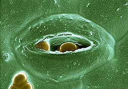
SEM of a Cryptosporidium oocyst in a spinach leaf stoma. Both images were taken for the ARS Environmental Microbiology and Food Safety Lab. Photo by ARS Electron and Confocal Microscopy Unit.
High-resolution images produced by U.S. Department of Agriculture (USDA) scientists at the Electron and Confocal Microscopy Unit (ECMU) in Beltsville, Md., are providing an unprecedented view into the extraordinary world of the unseen.
The images of specimens and samples magnified hundreds of thousands of times their original size have proven invaluable to researchers in describing new pest or pathogen species that pose a threat to U.S. agriculture. On the food safety front, use of the images has helped reveal the mechanisms by which certain bacteria, fungi and parasites infect fresh produce, according to Gary Bauchan, director of ECMU. The unit is part of the Agricultural Research Service (ARS), USDA's chief intramural scientific research agency.
ARS scientists at Beltsville routinely call upon the expertise of Bauchan and the microscopy team to image all manner of specimens and samples.
For example, when a plant pathologist at the ARS National Arboretum's Floral and Nursery Plants Research Unit contacted the team for assistance, the result was a three-dimensional image of how viruses spread from leaf veins to adjacent cells within a leaf. The image, which was generated using fluorescently tagged viruses and a Zeiss 710 confocal laser-scanning microscope (CLSM), illustrated how viruses are transferred within a leaf, and was the cover photograph in the November 2012 issue of the Journal of General Virology.
In addition to the CLSM, the research unit is equipped with a low-temperature scanning electron microscope (LTSEM), a variable-pressure scanning electron microscope, two transmission electron microscopes and a Hirox Digital video microscope. The microscopes are also equipped with digital cameras to speed the delivery of resulting images to researchers.
According to Bauchan, each microscope offers unique capabilities, but requires special handling to prepare specimens and samples prior to imaging. For systematic studies of mites, an LTSEM can be used. This necessitates flash-freezing a mite specimen (while still alive) at minus 321 degrees Fahrenheit and coating it with platinum. This freeze-frames the mite in time, allowing detailed examination of its microscopic features, behavior, and interaction with its immediate surroundings.
Read more about the ECMU and its contributions in the August 2013 issue of Agricultural Research magazine.
Científicos utilizan los "ojos" de alta tecnología para espiar a un mundo microscópico
Imágenes de alta resolución producidas por científicos del Servicio de Investigación Agrícola (ARS) en la Unidad de Microscopía Electrónica y Confocal (ECMU por sus siglas en inglés) mantenida por el ARS en Beltsville, Maryland, están proveyendo una vista sin precedente de un mundo extraordinario.
Las imágenes de especímenes y muestras ampliadas cientos de miles de veces han sido imprescindibles para los investigadores realizando estudios de nuevas especies de plagas o patógenos que representan una amenaza a la agricultura de EE.UU.
En el campo de la seguridad alimentaria, el uso de las imágenes ha ayudado a revelar los mecanismos usados por bacterias, hongos y parásitos para infectar las hortalizas, según Gary Bauchan, director de la ECMU. La unidad es parte del ARS, el cual es la agencia principal de investigaciones científicas del Departamento de Agricultura de EE.UU. (USDA por sus siglas en inglés).
Los otros científicos del ARS en Beltsville rutinariamente utilizan la pericia de Bauchan y sus colegas para crear imágenes de todos tipos de especímenes y muestras.
Por ejemplo, cuando un patólogo de plantas en la Unidad de Investigación de Plantas Florales y Plantas de Viveros mantenida por el ARS en el Arboreto Nacional de EE.UU. pidió ayuda del grupo, el resultado fue una imagen en tres dimensiones de cómo los virus se extienden de las venas de hojas a las células dentro de las hojas. Esta imagen, la cual fue generada utilizando los virus marcados con fluorescencia y un microscopio confocal Zeiss 710 de barrido laser (CLSM por sus siglas en inglés) demostró cómo los virus se mueven dentro de una hoja, y esta imagen fue representada en la portada de la revista 'Journal of General Virology' (Revista de Virología General) de noviembre del 2012.
Además del CLSM, los investigadores tienen un microscopio electrónico de barrido de baja temperatura (LTSEM por sus siglas en inglés), un microscopio electrónico de barrido de presión variable, dos microscopios electrónicos de transmisión y un microscopio de video Hirox Digital. Estos microscopios también tienen cámaras digitales para acelerar la entrega de las imágenes a los investigadores.
Según Bauchan, cada microscopio ofrece capacidades únicas, pero requiere el manejo especial para preparar especímenes y muestras antes de hacer las imágenes. Para los estudios sistemáticos de los ácaros, los investigadores usan el LTSEM. En este procedimiento, se tiene que congelar el espécimen del acara en temperaturas de menos 321 grados Fahrenheit y cubrir el ácaro con platino. Este proceso captura el ácaro precisamente y permite una inspección detallada de sus rasgos microscópicos, su comportamiento y su interacción con su ambiente.
Lea más sobre los investigadores de la ECMU y sus actividades en la revista 'Agricultural Research' de agosto del 2013.


