Nottingham, United Kingdom
September 18, 2014
A multidisciplinary team of scientists at The University of Nottingham are using some of the most advanced X-ray micro Computed Tomography (CT) scanners to learn how to design plant roots so they can interact better with soil and capture water and nutrients more efficiently. This non-invasive technology will help Nottingham unearth some of the answers to one of the biggest challenges facing the world today — global food security.
Malcolm Bennett, Professor of Plant Sciences, said: “For the first time in 10,000 years of plant breeding, we can see a plant’s root architecture directly in the soil, as it is in the field, and use this information to select the most efficient varieties for farmers to grow.”
The new Hounsfield Facility, on the University’s Sutton Bonington Campus, brings together specialists from the Schools of Biosciences, Computer Science, Mathematics and Engineering to delve into the ‘Rhizosphere’ — the thin layer of soil directly influenced by the proteins and sugars released by roots and inhabited by microorganisms that live off discarded plant cells.
The Nottingham scientists are using the
CT equipment and novel image analysis techniques to understand how roots of different crop varieties take up water and nitrogen. The aim is to find plants that use water and nutrients most efficiently to produce higher yields in more challenging conditions — such as drought and flooding.
Visualising plant root behaviour from seed to flowering
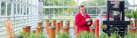
The Hounsfield Facility is equipped with three CT scanners — capable of imaging objects as fine as a soil particle or a root hair to a fully mature root system. It has a fully automated greenhouse which is manned by the laser guided robot, needed to feed the 1m long, 80kg samples to the largest scanner.
The research centre has been named after Sir Godfrey Hounsfield, the electrical engineer from Newark in Nottinghamshire, who shared the 1979 Nobel Prize in Medicine for his part in developing the diagnostic technique of X-ray Computed Tomography (CT).
With these scanners and specialised image analysis methods Nottingham researchers can image the structure of plant roots in a non-destructive way growing through soil throughout the life of the plant — from seed to flowering.
Imaging the hidden half of plants
Sacha Mooney, Professor of Soil Physics said: “We have finally overcome a major obstacle to our research. The opacity of soil prevented us from being able to see how roots actually grow in their natural environment. Using X-rays we can now ‘see-through’ the soil and visualise roots in 3D, offering new insights into the previously ‘hidden half’ of a plant. These new imaging technologies combined with biological resources have helped to create a world-leading facility with the tools that will radically improve our efforts to increase crop performance.”
There’s a small high resolution scanner for visualising fine details such as single roots, root hairs and the soil around them: a medium scale micro CT scanner to image an entire root system: and a large custom designed CT system to look at plants such as wheat throughout its whole growth cycle from seed to flowering — bringing the field closer to the lab than ever before.
X-ray CT produces a 3D image of the scanned sample. The RooTrak image analysis software, developed by experts in the University’s School of Computer Science, identifies root material within those images and builds a 3D model of the root system that can be viewed from any angle.
Tony Pridmore, Professor of Computer Science, said: “The problem with CT images is that roots and soil can appear very similar, and identical in some cases. RooTrak treats the 3D image as a set of 2D slices — a video — and tracks roots as they weave and branch through the soil. This allows it to adapt to the roots’ surroundings and extract root segments that can be stitched together to create a 3D model.”
The tracking method has recently been extended to allow RooTrak to separate one root system from another when multiple plants are grown in the same pot. This leads to 3D models showing the interactions between neighbouring, and even touching, root systems as they exist in an agricultural field competing for water and nutrients.
Research and commercial collaborations
The new centre has been funded by the European Research Council, BBSRC, the Wolfson Foundation and The University of Nottingham.
The scanners are also well suited to analysing a whole range of bio and non-biomaterials such as carbon fibres, food products, and electrical components. The team works with a wide range of local and national groups including Nottingham’s School of Veterinary Medicine and Science, the Faculty of Engineering, the British Geological Survey, several multinational food companies and even Michelin starred chefs!
Official opening
The new Hounsfield Facility will be officially opened on The University of Nottingham’s Sutton Bonington Campus by Professor Jackie Hunter, CE of the Biotechnology and Biological Sciences Research Council (BBSRC). Sir Godfrey Hounsfield’s niece will also attend representing the Hounsfield family.